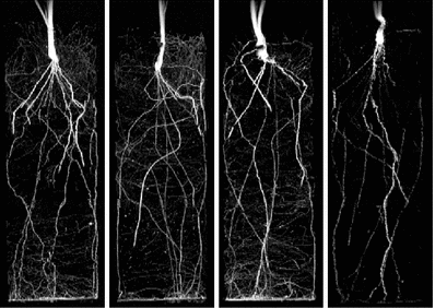
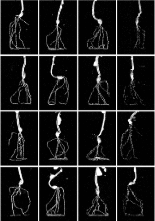
3D Magnetic resonance images of barley roots
IBG2 @ Forschungszentrum Juelich
Overview
| Organ | root-system |
|---|---|
| Number of images | 56 | Ground-truth | no |
| Other information |
Scientific article(s)
Description
Barley (Hordeum vulgare L. var Barke) plants (N=4) were cultivated in seven different substrates as detailed in the file ‘PlantIDs.xlsx’. Three weeks after sowing, the plant roots were imaged using MRI and subsequently harvested, scanned and analysed with WinRhizo. Both 3D MRI data and corresponding scanned images of the excavated roots are supplied. MRI acquisition parameters: 4.7T MRI equipped with a Varian console; Sequence: Spin Echo Multi-Slice (SEMS), TR=2850ms, Bandwidth BW=156kHz, Echo Time TE=9ms, two averages. Resolution: 0.5x0.5x1.0mm3. The 3D MRI data is supplied in the Nifti-file format, consisting of a header (*.hdr) and a data file (*.img). Furthermore, for each plant a maximum intensity projection is added as a preview image (*.tif).
Source: Dataset webpage